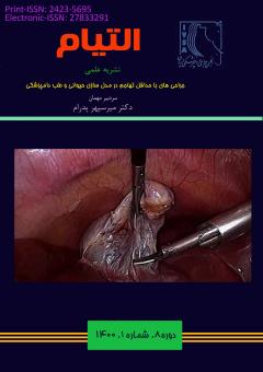
بررسی تاثیر انواع مختلف کانولا و همچنین اندازه ی اولیه ی برش, بر روی پوست و بافت های زیرین آن طی جراحی های لاپاروسکوپیک در مدل حیوانی سگ
محورهای موضوعی : علوم جراحی دامپزشکی شامل جراحی های بافت های سخت و نرم
مهدیه کاتبیان
1
*
,
محمد حجازی
2
![]() ,
روجا ابراهیمی
3
,
جلال رضایی
4
,
حسین مرجان مهر
5
,
حسین عاشق
6
,
فرناز محمودزادگان
7
,
حسام الدین اکبرین
8
,
روجا ابراهیمی
3
,
جلال رضایی
4
,
حسین مرجان مهر
5
,
حسین عاشق
6
,
فرناز محمودزادگان
7
,
حسام الدین اکبرین
8
![]()
1 - دانشجو، گروه جراحی و تصویربرداری تشخیصی، دانشکده دامپزشکی، دانشگاه تهران، تهران
2 - استادیار، گروه علوم درمانگاهی، دانشگاه آزاد اسلامی، تهران
3 - دانش آموخته، گروه جراحی و تصویربرداری تشخیصی، دانشکده دامپزشکی، دانشگاه تهران، تهران
4 - دانشیار، گروه علوم درمانگاهی، دانشکده دامپزشکی، دانشگاه تبریز، تبریز
5 - دانشیار، گروه پاتوبیولوژی، دانشکده دامپزشکی، دانشگاه تهران، تهران
6 - دانش آموخته، گروه جراحی و تصویربرداری تشخیصی، دانشکده دامپزشکی، دانشگاه تهران، تهران
7 - دانش آموخته، گروه جراحی و تصویربرداری تشخیصی، دانشکده دامپزشکی، دانشگاه تهران، تهران
8 - استادیار، گروه بهداشت مواد غذایی، دانشکده دامپزشکی، دانشگاه تهران، تهران
کلید واژه: لاپاروسکوپی , کانولا, سگ,
چکیده مقاله :
با وجود انجام مطالعات فراوان بر تاثیرات انواع تروکار بر التیام زخم در جراحی های لاپاروسکوپی, تا به حال تاثیر بدنه ی کانولا در هیچ مطالعهای مورد بررسی قرار نگرفته است. این مطالعه انجام شد تا مشخص شود که زخم حاصل از کدام نوع کانولا با التیام بهتری همراه است و اینکه آیا اندازهی برش محل پرت تاثیری بر پروسه التیام آن دارد؟ دراین مطالعه از ۵ سگ مادهی نژاد مخلوط استفاده شد. ۶ تروکار به دیواره شکم هر سگ وارد شد و گروههای مطالعه به این صورت تشکیل شدند: 10 ورود کانولای رزوه دار در برش ۷ میلی متری ( گروه A ) , 10ورود کانولای زروه دار در برش ۱۰ میلی متری (گروه B ) و 10 ورود کانولای صاف و برش ۱۰ میلی متری ( گروهC ). مقایسهی ماکروسکوپیک و هیسوپاتولوژیک گروه A و B نشان دهنده وسعت بالاتری از آسیب به لایهی پوستی و عضلانی شامل تغییرات دژنراتیو و نکروز و همچنین از دست رفتن بیشتر لایهی صفاتی در گروه A در مقایسهی B بود. مقایسهی هیستوپاتولوژیک دو گروه B و C نشان دهندهی خونریزی شدیدتری در لایه های درمان و تغییرات التهابی بیشتر در گروه B نسبت به C بود. این مطالعه نشان داد که برشهایی با طولی کمتر از قطر بدنهی خارجی کانولا تاثیری آسیب زننده بر بافت اطراف محل برش دارند. علاوه بر این مشخص شد کانولاهای زروه دار می توانند اثراتی منفی برای بافت اطراف خود داشته و نسبت به کانولاهای صاف آسیب زننده تر هستند.
Objective- While many of studies have evaluated effects of trocar on incision characteristics non has taken the design of the cannula into consideration. This study was conducted to figure out the type of cannula design which is associated with a better healing at the insertion site, and to investigate if the size of incision in the port site has an effect on the healing process. Procedure-6 trocars were inserted in each dog. five animals were used, allowing the total number of 10 insertions for 7 mm incisions and threaded cannula (group A), 10 insertions for 10 mm incision and threaded cannula (group B) and 10 for 10 mm incision and smooth cannula (Group C), which constituted 3 groups of study. Results-Macroscopic and Histopathology comparison between group A and group B revealed significantly higher degenerative changes and necrosis in the dermal and muscle layer and a higher loss of the peritoneal lining in group A than B. Hemorrhage in the dermal layer of the skin and acute inflammatory reaction was significantly higher in group B compared with C . Conclusions - This study showed that a smaller incision than the trocar’s external diameter has destructive effects on the tissues. Moreover, using a trocar with a threaded cannula can have harmful effects on the surrounding tissues and it is considered more destructive than a smooth cannula.
1. Bhoyrul S, Vierra MA, Nezhat CR, Krummel TM, Way LW. Trocar injuries in laparoscopic surgery. Journal of the American College of Surgeons. 2001;192(6):677-83.
2. Montz FJ, Holschneider CH, Munro MG. Incisional hernia following laparoscopy: a survey of the American Association of Gynecologic Laparoscopists. Obstet Gynecol. 1994;84(5):881-4.
3. Kadar N, Reich H, Liu CY, Manko GF, Gimpelson R. Incisional hernias after major laparoscopic gynecologic procedures. American journal of obstetrics and gynecology. 1993;168(5):1493-5.
4. McMurrick PJ, Polglase AL. Early incisional hernia after use of the 12 mm port for laparoscopic surgery. The Australian and New Zealand journal of surgery. 1993;63(7):574-5.
5. Tarnay CM, Glass KB, Munro MG. Incision characteristics associated with six laparoscopic trocar-cannula systems: a randomized, observer-blinded comparison. Obstet Gynecol. 1999;94(1):89-93.
6. Bohm B, Knigge M, Kraft M, Grundel K, Boenick U. Influence of different trocar tips on abdominal wall penetration during laparoscopy. Surgical endoscopy. 1998;12(12):1434-8.
7. Kolata RJ, Ransick M, Briggs L, Baum D. Comparison of wounds created by non-bladed trocars and pyramidal tip trocars in the pig. Journal of laparoendoscopic & advanced surgical techniques Part A. 1999;9(5):455-61.
8. McKay D, Blake G. Optimum Incision Length for Port Insertion in Laparoscopic Surgery. Annals of The Royal College of Surgeons of England. 2006;88(1):78-.
9. Kisiel A.,Single A.,Brisson B. “Maximizing the use of minimally invasive surgery in small animals:Laparascopy and Thoracoscopy.”Small animal veterinary rounds 1(8) (2012).
10. Munro MG, Tarnay CM. The impact of trocar-cannula design and simulated operative manipulation on incisional characteristics: a randomized trial. Obstet Gynecol. 2004;103(4):681-5.
11. Shafer DM, Khajanchee Y, Wong J, Swanstrom LL. Comparison of five different abdominal access trocar systems: analysis of insertion force, removal force, and defect size. Surgical innovation. 2006;13(3):183-9.
12. Zhao J, Liao D, McMahon BP, O’Donovan D, Schiretz R, Heninrich R, et al. Functional luminal imaging probe geometric and histomorphologic analysis of abdominal wall wound induced by different trocars in pigs. Surgical endoscopy. 2008;23(5):1004-12.

